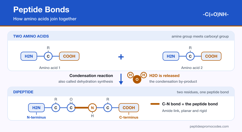
Almost everything interesting about peptides traces back to a single repeated connection. Strip away the sequences, the folding, and the biological activity, and what remains is one amino acid fastened to the next by a peptide bond. That bond decides how stable a chain is, what shape its backbone can take, and which conditions will eventually pull it apart.
This article works through the chemistry step by step: what the bond is, the reaction that builds it, why it counts as covalent, the properties that follow from its geometry, and the resonance effect that keeps it flat and rigid.
What Is a Peptide Bond?
A peptide bond is the chemical connection created between the carboxyl group of one molecule and the amino group of another. As the two groups react, a molecule of water leaves the reaction. Chemists call that process dehydration synthesis, or equivalently a condensation reaction.
By far the most common place this happens is between amino acids. The linkage that results is written CO-NH, and any molecule carrying that arrangement is an amide. The functional group at the heart of it — sometimes called the peptide group — is made of four atoms and is written -C(=O)NH-.
Everything built on this principle scales from there. Polypeptides and proteins are simply chains of amino acids fastened end to end by peptide bonds, one after another, which is why the bond turns up in essentially every discussion of what peptides are and what they do.
The two ends of a finished chain
Each bond consumes one amino acid’s carboxyl group and the next amino acid’s amino group. Once a chain is assembled, one end still carries a free amino group and the opposite end still carries a free carboxyl group. The free-amine end is the N-terminus, the free-acid end is the C-terminus, and sequences are conventionally read and written starting from the N-terminus. That written order of residues is the chain’s primary structure.
How Do Peptide Bonds Form?
Two amino acids join when the carboxylic acid group on one reacts with the amine group on the other. The simplest possible version of this reaction involves exactly two amino acids and produces a dipeptide, the smallest peptide there is. Repeat the step and the chain keeps growing.
Chain length is what separates the vocabulary used across the field, although the cut-offs are loose conventions rather than fixed rules. The IUPAC-IUBMB nomenclature puts them roughly here:
| Number of amino acids | What the chain is called |
|---|---|
| Fewer than about 10 to 20 | Oligopeptide |
| More than about 10 to 20 | Polypeptide |
| More than about 50, in a specific sequence | Usually called a protein |
The same recommendations note that authors differ greatly on where “protein” begins, and a common shorthand simply calls any chain of up to about 50 residues a peptide. Those thresholds are the basis for the wider comparison between peptides and proteins, but the bond doing the joining is the same one at every length.
Breaking the bond again
The reverse of formation is hydrolysis. Exposure to water breaks a peptide bond, splitting the chain back apart — the same water that was expelled during synthesis is taken up again during cleavage.
That does not mean peptide chains fall apart on contact with moisture. Peptide bonds are metastable: hydrolysis is energetically downhill, but without a catalyst it is extremely slow. At neutral pH, the half-life of an uncatalyzed amide bond has been measured at about 267 years. The hydrolysis reaction yields only about 10 kJ/mol (roughly 2.4 kcal/mol) of free energy.
| Property | Formation (condensation) | Breakdown (hydrolysis) |
|---|---|---|
| Water | One molecule released | One molecule consumed |
| Effect on the chain | Bond created, chain extended | Bond cleaved, chain shortened |
| Rate | Catalyzed by the ribosome in living cells | Very slow without a catalyst; the bond is metastable |
| Energy | Requires input | Yields roughly 10 kJ/mol of free energy |
Living organisms handle both directions with catalysts, but not the same ones. Peptide bonds are made on the ribosome, whose large subunit drives peptide bond formation at around 20 bonds per second, while a separate family of enzymes, the proteases, hydrolyze them. Between them, that is how tissue is built, remodeled, and digested. That biological route is worth contrasting with the laboratory methods used to make peptides deliberately.
Spotting peptide bonds in the lab
Peptide bonds absorb far-ultraviolet light, most strongly near 190 nm. Because measuring that low is technically awkward, labs often read absorbance a little higher, at 205 nm, where the signal still comes mainly from the peptide backbone. That absorption is a practical handle for detecting and measuring peptides.
Where you encounter peptide bonds
Because the bond is so general, the molecules built from it span an unusually wide range of applications, including:
- Hormones: signaling molecules that coordinate processes between tissues.
- Antibiotics: peptide-based compounds used against microbial targets.
- Anti-aging products: cosmetic and topical formulations built on short peptide chains.
- Anticancer medicines: therapeutic agents assembled from peptide sequences.
- Weight-loss supplements: products marketed around peptide activity.
- Neurotransmitters: peptide messengers operating in the nervous system.
Many of the compounds sold under the research peptides label belong to these same categories, and every one of them depends on peptide bonds holding the sequence together.
Are Peptide Bonds Covalent?
Yes. The link forms through the sharing of electrons between a carbon atom and a nitrogen atom, and electron sharing is the defining characteristic of a covalent bond.
More precisely, the peptide bond is a covalent bond of the amide type. It connects consecutive alpha-amino acids along a chain, tying C-1 of one residue (its carboxyl carbon) to N-2 of the next (its alpha-amino nitrogen).
What Are the Characteristics of Peptide Bonds?
The bond’s behavior is best understood as a short list of properties that follow directly from its structure:
- Strength: peptide bonds are strong, and they carry partial double-bond character rather than behaving as plain single bonds.
- Resistance to heat and salt: high salt concentrations and heating do not readily break them.
- What does break them: a strong acid or base, applied at high temperature over an extended period, will cleave the bond. Amino acid analysis relies on exactly this: one published protocol hydrolyzes peptides in 6 N hydrochloric acid at 110°C for 18 hours.
- Enzymatic cleavage: specific enzymes, digestive enzymes among them, break peptide bonds under far gentler conditions.
- Stiff and planar geometry: the rigid, flat arrangement of the bond is what preserves the structural integrity of a protein.
- Polarity: the bond contains a slightly positive polar hydrogen on the amide nitrogen (N-H) and a partly negative polar oxygen on the carbonyl group (C=O).
That combination — chemically tough, but selectively cleavable by enzymes — is precisely what a biological polymer needs. Chains hold their shape under everyday conditions while remaining available for controlled breakdown when the organism requires it.
The Structure of the Peptide Bond
The structural description of the peptide bond was established by Linus Pauling and Robert Corey. Pauling derived it from resonance theory, and careful experimental studies by Corey and his coworkers verified it.
What they described is a planar, rigid arrangement that exhibits partial double-bond character, and in proteins it is almost always trans. The coplanarity comes from resonance: the amide nitrogen partly shares its lone pair of electrons with the carbonyl group.
The practical consequences of that arrangement are:
- A single plane: the carbon, hydrogen, nitrogen, and oxygen atoms of the peptide group all sit in the same plane, together with the alpha carbons on either side, six atoms in all.
- A trans orientation: the carbonyl oxygen and the amide hydrogen point in opposite directions, trans to one another. The cis form is rare: about 0.03% of ordinary peptide bonds are cis, rising to about 5% for bonds that precede a proline.
Because a whole chain is a series of these flat, fixed units, the backbone’s flexibility is restricted to the bonds on either side of each peptide group rather than the peptide bond itself.
The Polarity of the Peptide Bond
Under normal circumstances, a single bond between the carbonyl carbon and the amide nitrogen would permit free rotation. The peptide bond does not behave that way, and resonance is the reason.
The nitrogen atom carries a lone pair of electrons positioned close to the carbon-oxygen bond. That proximity makes a second resonance structure possible: one in which a double bond exists between the carbon and the nitrogen, leaving the nitrogen with a positive charge and the oxygen with a negative charge.
The existence of that resonance form prevents rotation around the peptide bond. The true structure of the bond is neither resonance form on its own but a weighted combination of the two, and the result is a bond with approximately 40% double-bond character. Pauling’s estimate was backed by the bond’s measured length of 1.32 Å.
That figure explains the rest of the bond’s behavior. The rigidity described in the previous sections is a direct consequence of this partial double-bond character, and so is the fixed planar geometry that the surrounding atoms are forced to adopt.
Why the Bond Matters in Practice
Reduced to essentials, the peptide bond does three jobs at once. It joins amino acids into a defined sequence, it holds that sequence together under ordinary conditions of heat and salt, and it stays vulnerable to the specific enzymes that need to take chains apart on demand.
Every property that makes peptides useful — the ordered sequence, the stable backbone, the predictable shape — sits downstream of this one amide linkage and the roughly 40% double-bond character that keeps it flat.
Understanding peptide bond chemistry helps you read product and study descriptions with a clearer eye, but it is not a substitute for medical guidance. Consult a qualified healthcare professional before beginning any peptide regimen or therapy.
Sources
- IUPAC-IUBMB Joint Commission on Biochemical Nomenclature: Nomenclature and Symbolism for Amino Acids and Peptides, 3AA-11 to 3AA-13 (Recommendations 1983): the definition of a peptide bond, the C-1 to N-2 link, loss of water, N- and C-termini, and the loose length cut-offs for oligopeptides, polypeptides and proteins.
- Linus Pauling: Modern Structural Chemistry (Nobel Lecture, 1954): amide resonance, 40% C-N double-bond character, the planar six-atom group, the 1.32 Å bond length and Corey’s experimental work.
- Mahesh, Tang and Raj: Amide Bond Activation of Biological Molecules (Molecules, 2018): why amide bonds are so stable, how resonance makes them planar and hinders rotation, and the 267-year half-life at pH 7.
- Orgel: The Implausibility of Metabolic Cycles on the Prebiotic Earth (PLoS Biology, 2008): a free energy of hydrolysis of about 2.4 kcal/mol per peptide bond, and why forming the bond needs an energy source.
- The Nobel Prize in Chemistry 2009: Popular Information: the ribosome’s large subunit forming peptide bonds at about 20 per second.
- López-Otín and Bond: Proteases, Multifunctional Enzymes in Life and Disease (J Biol Chem, 2008): proteases as the enzymes that hydrolyze peptide bonds.
- Anthis and Clore: Sequence-Specific Determination of Protein and Peptide Concentrations by Absorbance at 205 nm (Protein Science, 2013): far-UV absorbance peaking near 190 nm, with absorbance at 205 nm coming mainly from the peptide backbone.
- McCarthy et al.: Reference Standards to Support Quality of Synthetic Peptide Therapeutics (Pharmaceutical Research, 2023): acid hydrolysis of peptides in 6 N HCl at 110°C for amino acid analysis.
- Exarchos et al.: Detection of Discriminative Sequence Patterns in the Neighborhood of Proline cis Peptide Bonds and Their Functional Annotation (BMC Bioinformatics, 2009): most peptide bonds are trans, with about 0.03% of non-proline and 5.2% of proline-preceding bonds found in cis.